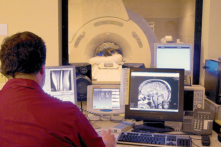
Medical imaging refers to several different technologies that are used to view parts of the human body in order to diagnose, monitor, and treat medical conditions. Each type of medical imaging gives us different information about the area of the body being treated, which can be related to injury, disease, or determining the effectiveness of medical treatment.
Whether you have experienced an injury, are suffering from chronic pain, or are facing another medical condition, medical imaging allows your doctor to determine what is going on inside your body and recommend the proper treatment.
Common types of medical imaging
X-Ray
X-ray technology is one of the oldest and most common forms of medical imaging used by doctors. X-rays are a form of electromagnetic radiation that send small waves through the body, which are then absorbed in different amounts depending on the materials they pass through. Dense materials like bone or metal will show up white on x-rays, while air in your lungs would show up black. Materials like fat and muscle often appear as shades of grey.
Typically used to evaluate:
- Broken bones
- Arthritis in the joints
- Blood vessels
- Dental issues
- Lungs
- Mammography
- Foreign objects (such as something swallowed by a child)
Ultrasound
Medical ultrasonography uses high frequency sound waves to create images of various organs and tissues inside the body. An ultrasound machine sends sound waves into the body and converts the returning sound echoes into an image. Ultrasound technology can also produce sounds related to blood flow, which can be helpful to medical professionals depending on your condition.
Typically used to evaluate:
- Pregnancy
- Conditions of the heart and blood vessels
- Pelvis and abdomen
- Pain, swelling, and/or infection
- Help guide certain procedures such as injections or biopsies
CT or CAT Scan
Computed tomography (CT), sometimes called computed axial tomography (CAT), uses specialized x-ray technology and computing algorithms to create images of the inside of the body. Multiple x-ray projections are taken from different angles to produce detailed, 360-degree cross-sectional images (tomograms). CT images allow medical professionals to view very precise, 3-D views of certain parts of the body, which can be extremely helpful for diagnostic purposes.
Typically used to evaluate:
- Imaging bone, soft tissue, and blood vessels at the same time
- Lung and chest issues
- Cancer and tumors
- Bone injuries
- Traumatic injuries
- Cardiac conditions
- Organs in the pelvis and abdomen
MRI
Magnetic Resonance Imaging (MRI) uses radio waves (radiofrequency energy) and a strong magnetic field to produce accurate images of internal body structures. During an MRI, an electric current is passed through coiled wires to create a temporary magnetic field in the patient’s body. Radio waves are sent and received by a machine, and the signals are used to create digital images of the scanned area of the body. For some MRI exams, intravenous (IV) drugs are used to change the contrast of the images produced. MRI’s are an essential tool in diagnosing and treating musculoskeletal and neurological conditions.
Typically used to evaluate:
- Tendon and ligament tears
- Spinal conditions and injuries
- Bones and joints
- Imaging organs, soft tissue, and other internal structures
- Blood vessels
Who should I talk to about medical imaging?
Our highly trained and compassionate radiologists and medical imaging specialists have the experience to help you, no matter what injury or condition you may be experiencing. They work with you and your doctor to ensure the highest quality images and experience.
The Center offers digital x-rays at our Neff Road clinic, Redmond clinic, and Shevlin Health and Wellness Center clinic. Magnetic Resonance Imaging (MRI) is available at our Neff Road clinic only.


