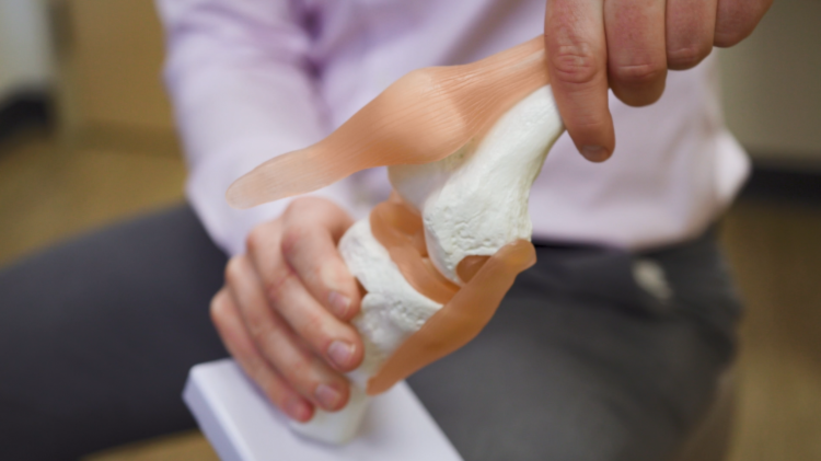
The knee is a complex joint with many different components. Bones, tendons, ligaments, and cartilage all work together to allow the joint to move and bend smoothly. It is also one of the largest joints in your body, making it vulnerable to a variety of injuries. Knee pain can be debilitating and impact your ability to do even the simplest everyday activities. Some of the most common knee injuries include overuse injuries, degenerative conditions, dislocations, sprains, fractures, and ligament tears.
Knee ligament sprains and tears are the most common knee injury, often seen in individuals who participate in sports from football to soccer to skiing. Each year in the United States approximately 75,000 people will experience an MCL injury and up to 200,000 will experience an ACL injury.
The anatomy of the knee
The knee is a hinge joint created by the femur (thighbone) and the tibia (shinbone). The patella (kneecap) is the third bone present that covers and protects the joint. In addition to these bones, cartilage, ligaments, and tendons are all present within the knee to help the joint function properly.
Ligaments are the tissues that connect bones to other bones. There are four main ligaments in your knee that hold the bones of the joint together and provide stability:
- Anterior cruciate ligament (ACL)
- Lateral collateral ligament (LCL)
- Medial collateral ligament (MCL)
- Posterior cruciate ligament (PCL)
There are collateral and cruciate ligaments. Collateral ligaments (lateral collateral ligament and medial collateral ligament) are found on each side of your knee and control sideways movement, while cruciate ligaments (anterior cruciate ligament and posterior cruciate ligament) are found inside your knee joint and control backward and forward movement.
Understanding ligaments
Your ligaments are made up of fibrous soft tissues that hold your bones and joints together. Picture your ligaments as strong ropes – a small cut in the rope will make it less stable as all of the fibers become loose. This is similar to a tear in one of your ligaments, which occurs when the fibers are partially or fully torn away from each other. As the tissues in your ligaments expand and retract to stabilize your joints, the fibers can tear from certain unnatural movements like a fall or collision.
What is an ACL Tear?
The ACL is located inside of the knee joint and connects the top front of the tibia (shinbone) to the bottom back of the femur (thighbone). Together, the ACL and PCL cross each other to form an “X” inside of your knee. The ACL prevents the tibia from sliding too far forward relative to the femur and is important for rotational stability. ACL injuries often occur during sports activities that involve frequent pivoting or cutting, like football, soccer, and basketball. Quickly changing direction or landing from a jump incorrectly can also result in an ACL injury.
What is a MCL Tear?
The MCL is located on the inner side of your knee (connecting the bottom of the femur to the top of the tibia) and works to control sideways motion. Injuries to the MCL are usually caused by contact injuries to the outside of the joint that push the knee inward. Football, hockey, and other high contact sports are often responsible for MCL injuries.
It is not always the case, but ACL injuries are generally more complex to treat than MCL injuries, which sometimes heal on their own.
ACL and MCL injury symptoms
Injured ligaments are rated on a scale of one to three. Grade 1 means mild damage where the ligament has been slightly stretched, Grade 2 means a stretch to the point where the ligament is loose (partial tear), and Grade 3 means the ligament has been torn into two pieces.
ACL tear symptoms
- Pain and tenderness with swelling
- A “popping” noise or feeling of the knee giving out
- Difficulty and discomfort while walking
- Loss of full range of motion
MCL tear symptoms
- Pain and swelling specifically on the sides of the knee
- Instability or feeling of the knee giving out
In both cases, you are likely to experience pain and a significant instability of the injured knee. You may still be able to bear weight after a torn ACL, or you may not be able to depending on the severity of your injury. Most people are still able to walk and bear weight after an MCL tear.
ACL and MCL diagnosis
Most ligament injuries can be diagnosed through a physical examination of the knee where your doctor will compare the structure of your injured knee to your non-injured knee. Your doctor may or may not order an x-ray or MRI in order to determine whether the injury is associated with a broken bone or to get a better picture of the soft tissue ligaments.
ACL and MCL treatment
A MCL injury rarely requires surgery and, in time, will heal on their own given proper care. Icing, bracing, and physical therapy are all typical in an MCL tear treatment plan. Icing your injury is extremely important to the healing process and should be done for 15 to 20 minutes at a time with at least one hour between icing sessions.
It is important for your knee to be protected from the same sideways force that caused the injury, so your doctor may recommend a brace or even crutches to support the knee. You may need to adjust your daily activities to avoid bearing weight or risky movements. Strengthening exercises and physical therapy can help to restore function and strengthen the muscles that support the knee joint.
Your doctor may recommend nonsurgical treatment like bracing or physical therapy for an ACL injury, but a torn ACL will not repair itself. Sometimes patients who are elderly or who have a very low activity level and still have overall stability of the knee will not require surgery and can manage their injury with other treatment.
Surgical treatment of a torn ACL usually involves arthroscopy, where a small camera is inserted into the knee through a small incision and connected to a video monitor in the operating room. Your surgeon will use the camera to repair the damaged ACL with tissue from another part of the body – generally a tendon from the knee or hamstring, or from a cadaver. Choosing to use your own tissue or a cadaveric tissue graft is something you should discuss with your orthopedic surgeon, as there are pros and cons to each option. After the torn ACL is replaced with new tissue, it will develop into a ligament over time.
ACL and MCL recovery process
Once your range of motion returns to normal and you are able to walk and bear weight without a limp, your doctor will usually allow a gradual and progressive return to activities. This might look like starting with jogging and slowly progressing to sprinting over time. You may need to continue to wear a knee brace during physical activity depending on the severity of your injury.
ACL reconstruction surgery has a very high success rate. Your ACL rehabilitation plan is likely to include physical therapy, starting with a focus on returning motion to the joint and followed by strengthening exercises to protect your new ligament. After these steps have taken place, then you will follow a functional return to your normal physical activities. Generally, patients may return to sports when there is no longer any pain or swelling, typically a 4-6 month range. Recovery after surgery varies from patient to patient. Recovery time takes at least six months to fully incorporate the new ACL into your body.
ACL and MCL injuries are extremely common and often quite painful. The best thing you can do for a speedy recovery is to visit your doctor immediately following a knee injury to develop a treatment plan that works for you and your lifestyle.
If you’re looking for more tips and information on living a healthy and pain free life, join our monthly newsletter. Link to sign up is below.


